Arthroscopy is a closed surgery method used in the treatment of joint problems, which does not require to make large incisions, and allows the diagnosis and treatment of the joint by camera imaging. Arthroscopy, which is the most commonly used treatment method in the orthopedic field, is based on the principle of sending camera and auxiliary treatment tools through holes of approximately 0.5 cm and monitoring the damaged area from an external monitor. The images taken with the tiny camera can be enlarged to see the inside of the joint to the finest detail.
Arthroscopy can be used in all joints. The most commonly used one is knee arthroscopy. Shoulder arthroscopy is the second. Hip arthroscopy, ankle arthroscopy, and elbow arthroscopy are also preferred methods. Arthroscopy could not be done very frequently in the past because of the problem of vascular and neural networks in the elbow area and problems in the entrances and camera placement. However, in recent years, with the increasing experience in closed systems for the elbow joint, the feasibility of arthroscopic procedures has increased considerably.
What is Elbow Arthroscopy?
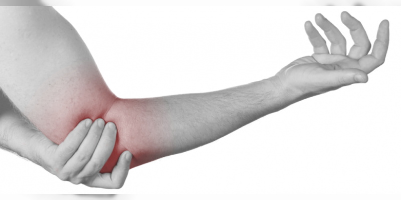
The elbow joint is more complex than the other joints. It is an important area that provides the opening, bending and rotation movements of the arm. Cartilage damage and osteoarthritis may occur on the elbow due to trauma or without trauma. Treatment may be performed by applying elbow arthroscopy in cases of jamming sensation, movement restriction, rheumatic diseases, calcification, and joint mouse. With arthroscopy, the soft tissues are not damaged and the possible movement limitations that occur later are eliminated. Joint mouse removal, calcification, soft tissue damage removal, joint capsule loosening, and synovectomy can be performed with the camera system. Since the scar is not large, the pain is less and the recovery process is shorter
Followings can be performed for elbow arthroscopy:
- Intraarticular detailed examination
- Ulnar nerve compression
- Cartilage repair
- Fracture treatment
- Repair of protruding bone
- Treatment of tendon ruptures
- Tennis and golfer’s elbow
How is Arthroscopy carried out?
Arthroscopy is carried out under general or local anesthesia. Approximately 0.5 cm 2 or 3 holes are made on the elbow joint and a micro camera (with fiberoptic light) and auxiliary treatment devices are inserted. The surgeon performs the repair by monitoring the camera’s data from the opposite monitor. A few parts can be intervened at the same time. During the procedure, photos and videos can be taken from within the joint. Since the joints are small in elbow, wrist and ankle arthroscopy, different (smaller) arthroscopes are used compared to those in the knee, shoulder, and hip.
Return to normal life after arthroscopy is much faster than open surgery. Because the incisions are small, the recovery process is painless, fast and comfortable.
Advantages of Elbow Arthroscopy
The patient heals faster, is discharged on the same day or the next day, returns to normal life faster. The scar disappears quickly, it is aesthetic. Less tissue damage occurs, more natural preservation of joints. The pain is less. Lower risk of complication. Lower risk of infection.


